


Medical Equipment Manufacturer
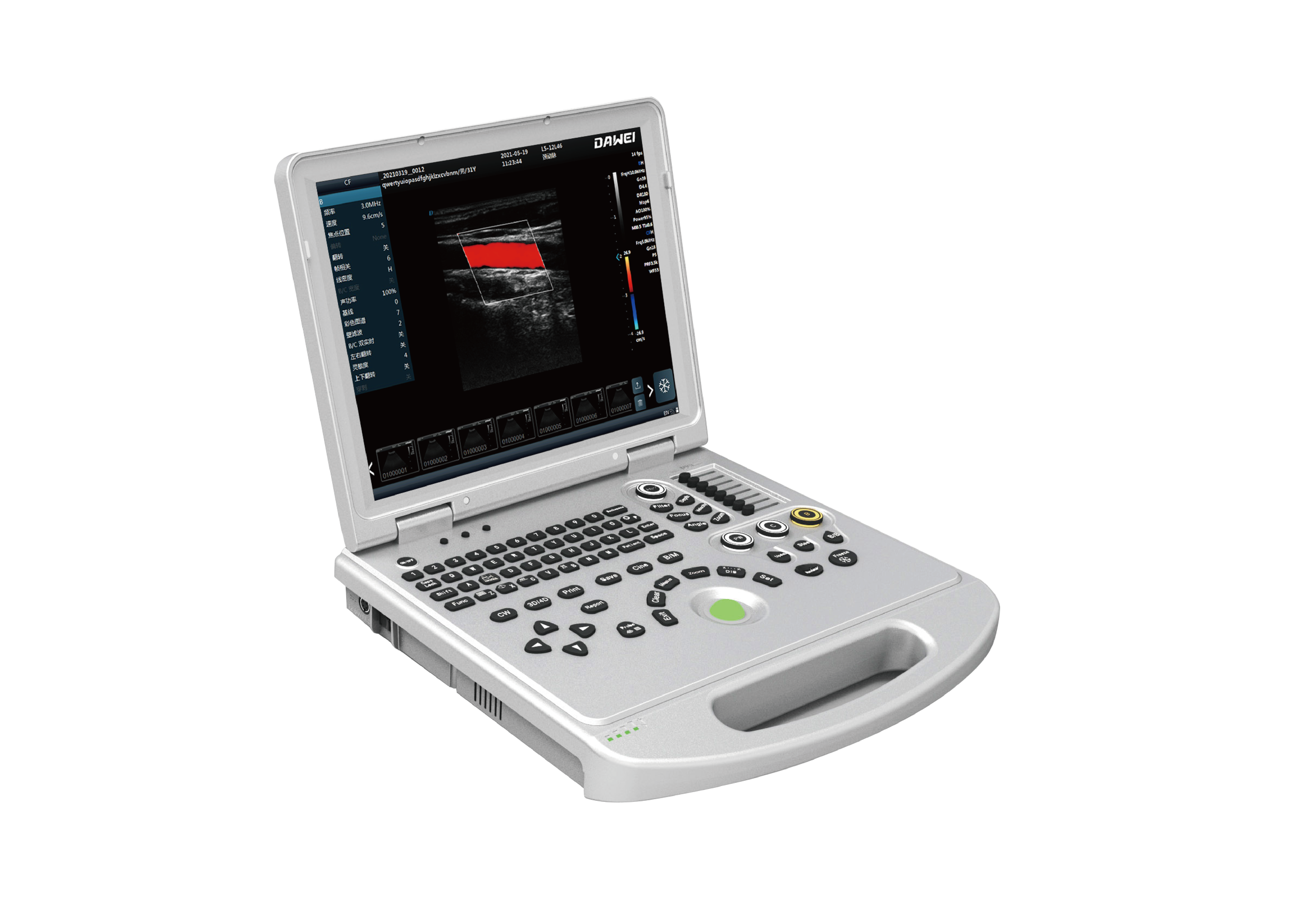
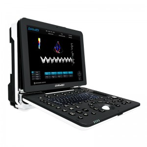
DW-L5(DW-PF522) color doppler ultrasound machine
-
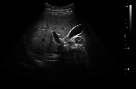
Micron imaging technology
Micron imaging technology, real-time tracking of specific signals at the edges of different tissues, to achieve edge enhancement, and monitor each pixel at the same time; optimize the internal signal of the organization and perfectly integrate the edge information and the internal pixel information of the organization to restore the real and delicate, excellent level contrast Two-dimensional image.
-
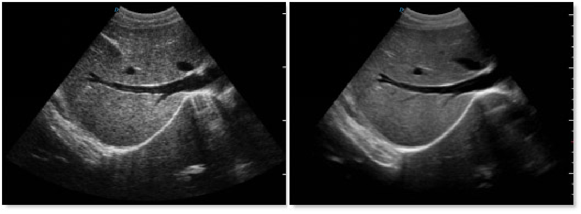
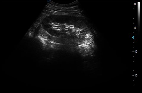
Harmonic imaging technology(THI)
It improves image clarity by improving tissue contrast resolution, spatial resolution, and eliminating near-field artifacts. It is
mainly used for the diagnosis of cardiovascular and abdominal diseases. It plays an important role in evaluating the lesion area and boundary division of patients with imaging difficulties. The technology has been fully approved by clinicians. Harmonic technology retains the second harmonic signal to
the greatest extent on the basis of removing the fundamental signal, which increases the signal strength by more than 30% compared with the traditional signal processing, reduces noise and artifacts, and improves the contrast resolution of
tissue images.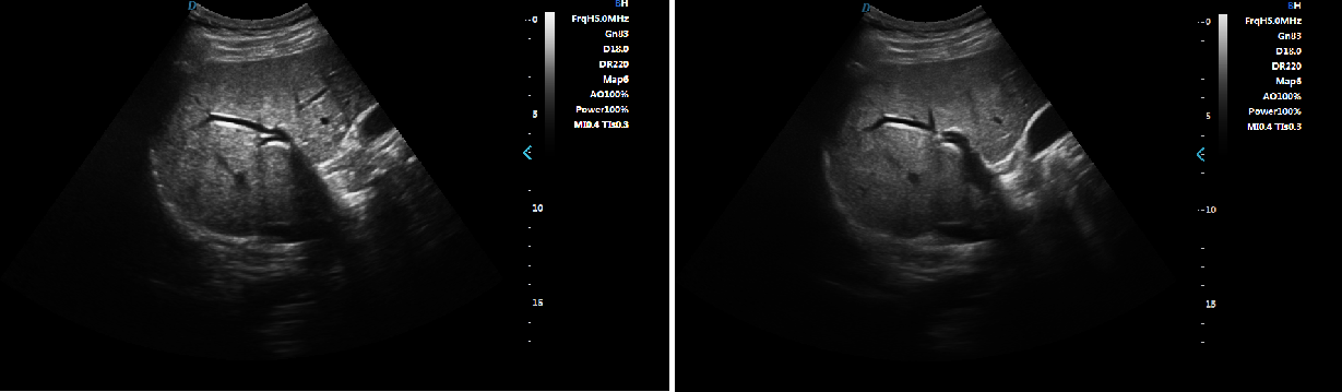
-
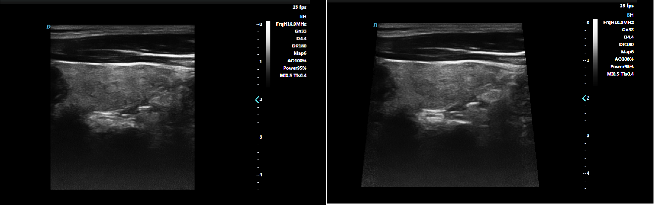
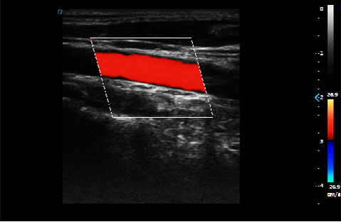
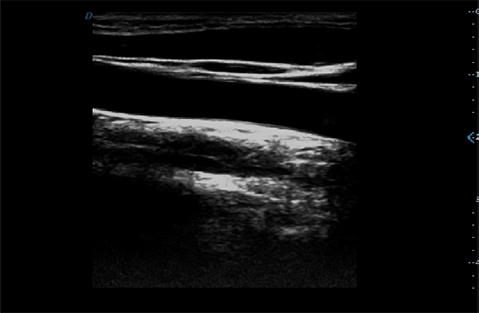
Trapezoid imaging
Trapezoid imaging is a kind of expanded imaging, which is transformed into a trapezoid on the basis of the original rectangle, and the left and right sides are expanded to a certain extent, achieving a wider field of view. The principle of ultrasound imaging is to scan the human body with ultrasonic sound beams, and obtain images of internal organs by receiving and processing the reflected signals.
-
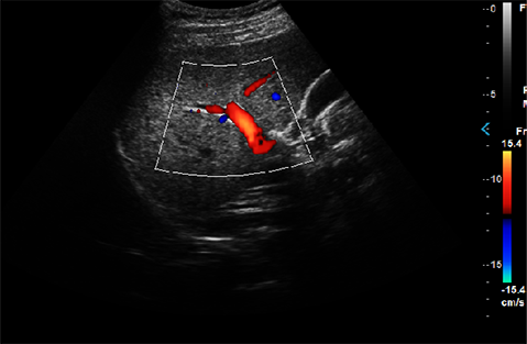
Automatic spectrum tracking measurement technology
Ultrasound Doppler technology is used in the ultrasound system for examining the heart and arteries and veins. It is necessary to extract relevant parameters from the Doppler spectrogram to evaluate the hemodynamic status of the heart and blood vessels. The disadvantage of manual detection is that the operator's marking of the peak velocity is
relatively monotonous and time-consuming, with poor repeatability and low estimation accuracy; and during the detection, in order to mark the peak velocity, the operator needs to interrupt the acquisition of Doppler signals, which makes it impossible to estimate in real time. This host contains an automatic envelope detection module, which can automatically track the time-related changes of the peak blood flow velocity and average velocity, and display them in real time on the Doppler spectrogram.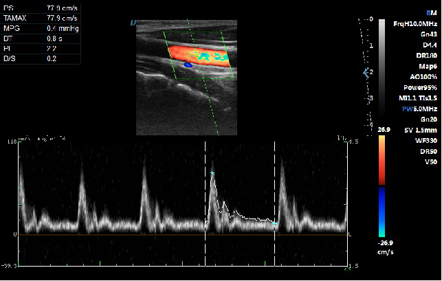

-
Optional Accessories
• Yellow dongle workstation:
(Direct patient file management, support image dynamic and static storage.)
• Foot switch.
• Puncture frame.
• Video printer and printer holder. -
Probe
• Convex probe
• Micro-convex probe
• Linear probe
• Trans-rectal probe
• Trans-vaginal probe
• Phased array probe
-
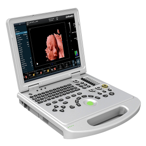
DW-L50(L5PRO) 3D/4D/5D Portable medical echo ul...
Perfect Obstetric Assistantmore>> -
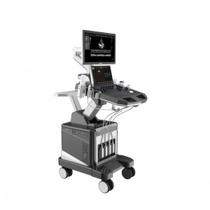
DW-T8 powerful echo ultrasound professional 4d ...
Comprehensive diagnostic solutions: rich measurement software can quickly and conveniently solve the needs of abdominal, peripheral vascular, gynecology, obstetrics, newborn, skeletal muscle, heart, and other clinical diagnoses.more>> -
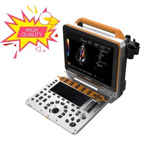
DW-P8(PF582) portable 4D cardiovascular ultraso...
From obese to Thin, Neonate to geriatric, with Dawei 4.0 ultrasound systemmore>> -
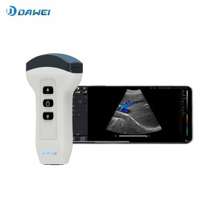
Wireless Handheld Ultrasound Scanner
The wireless handheld ultrasound system enables the flexible Performance of body scanning and delivers crystal-clear images.more>> -
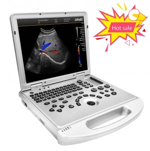
DW-L3 portable medical color Doppler ultrasound...
Perfect professional measurement package (abdomen, urology, etc.) to effectively solve the basic layer of medical needs.more>> -
DW-P60(P8Lite) Best Portable medical cardiac ul...
From obese to Thin, Neonate to geriatric, with Dawei V4.0S ultrasound systemmore>>